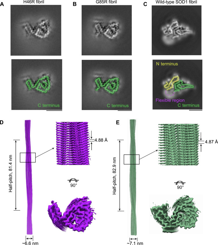
Fig. 1. Comparison of the cryo-EM structures of the H46R fibril and the G85R fibril.
(A and B) Cross-sectional view of the 3D map of the H46R fibril (A) or the G85R fibril (B) showing a protofilament comprising a C-terminal segment (green). (C) Cross-sectional view of the 3D map of the wild-type SOD1 fibril, however, showing a protofilament comprising not only a C-terminal segment (green) but also an N-terminal segment (yellow) with an unstructured flexible fragment (magenta dashed line) (43). Scale bars, 5 nm. For full clarity, we false color the equivalent regions in (A) to (C). (D and E) 3D map of the H46R fibril (D) or the G85R fibril (E) showing a single protofilament [in purple for (D) and green for (E)] intertwined into a left-handed helix, with a fibril core width of ~6.6 nm (D) or ~7.1 nm (E) and a half-helical pitch of 81.4 nm (D) or 82.9 nm (E) (left). Enlarged section of the H46R fibril (D) or the G85R fibril (E) showing a side view of the density map (top right). Close-up view of the density map on the left showing that the subunit in a protofilament stacks along the fibril axis with a helical rise of 4.88 Å (D) or 4.87 Å (E) (top right). Top view of the density map of the H46R fibril (D) or the G85R fibril (E) (bottom right).

