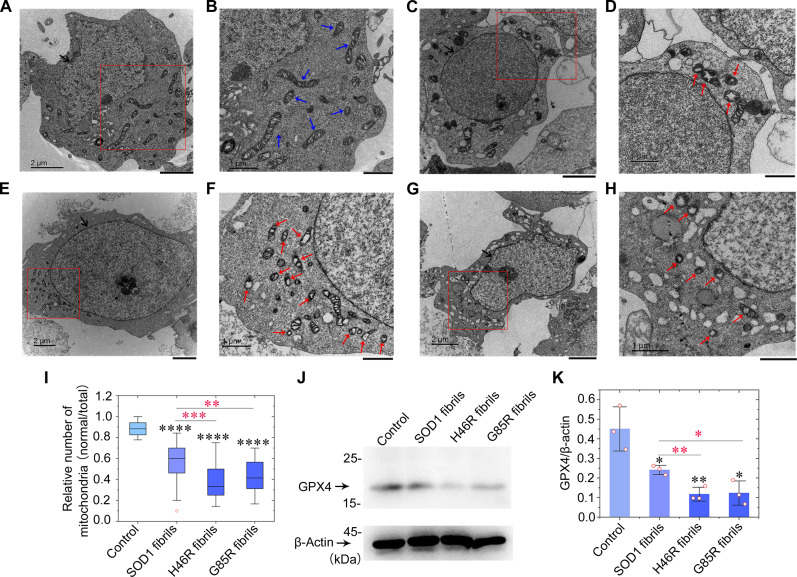
Fig. 5. Fibril seeds from H46R and G85R cause severe mitochondrial impairment and induce ferroptosis in neuronal cells.
(A to H) SH-SY5Y cells were cultured for 1 day and then incubated with 0 μM SOD1 fibril seeds (A and B), 10 μM wild-type SOD1 fibril seeds (C and D), 10 μM H46R fibril seeds (E and F), and 10 μM G85R fibril seeds (G and H), respectively, for 3 days. Nuclei and normal mitochondria in SH-SY5Y cells are highlighted using black arrows (A, C, E, and G) and blue arrows (B), respectively. Abnormal mitochondria with morphological features of ferroptosis or mitochondrial vacuolization are highlighted by red arrows (D, F, and H). Samples were negatively stained using 2% uranyl acetate and lead citrate. Scale bars, 2 μm (A, C, E, and G) and 1 μm (B, D, F, and H). (I) Box plot analyzing the relative number of mitochondria (normal/total) in SH-SY5Y cells treated with SOD1 fibril seeds and showing the quantification of TEM images in n = 30 SH-SY5Y cells examined over three independent experiments. (J) Western blot for GPX4 in the cell lysates from SH-SY5Y cells incubated with fibril seeds from H46R and G85R, compared with those incubated with wild-type SOD1 fibril seeds. β-Actin served as the protein loading control. (K) The relative amount of GPX4 in the above cell lines (open red circles shown in scatter plots) was determined as a ratio of the density of GPX4 band over the density of β-actin band in cell lysates and expressed as the means ± SD (with error bars) of values obtained in three independent experiments.

