Fig. 1.
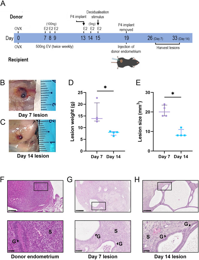
Development of endometriosis lesions in mice. (A) A modified version of a menstrual mouse model of endometriosis was used to establish subcutaneous endometriosis-like lesions in recipient mice. E2, beta-estradiol; EV, estradiol valerate; OVX, ovariectomy surgery; P4, progesterone. (B,C) Representative images of lesions at Day (D)7 (B) and D14 (C). (D,E) Lesions were excised, weighed (D) and measured (E). (F-H) Representative photomicrographs of Hematoxylin and Eosin (H&E)-stained sections from donor decidualized endometrium (F), D7 lesions (G) and D14 lesions (H). Top row: 10× magnification; bottom row: 40× magnification. Scale bars: 50 μm. G, glands; S, stroma. Data are presented as median±interquartile range, with each symbol representative of a single lesion in one mouse used for RNA sequencing (RNA-seq). Statistical analysis was performed using the Mann–Whitney test (*P<0.05).

