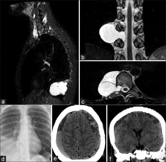
Figure 1.

Sagittal (a), coronal (b), and axial (c) T2-weighted magnetic resonance images depicting the right thoracic meningocele located at the T9-T11 level. Postoperative (thoracotomy) chest radiography (d) shows the pleural effusion along with the cyst residual. Axial computed tomography (CT) image (e), and coronal CT multiplanar reconstruction (f) demonstrate bilateral subdural hematoma secondary to intracranial hypotension
