Figure 2.

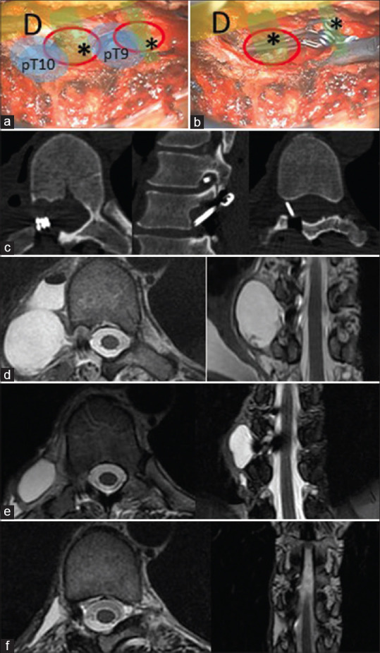
Intraoperative images after isolation (a) and clipping (b) of cyst’s neck. D: dural sac; pT9: T9 pedicle; pT10:T10 pedicle; *cyst’s caudal neck; *cyst’s cranial neck. Postoperative (laminectomy and cyst exclusion) images: bone window axial, and sagittal reconstruction computed tomography images (c) show cyst’s necks clipping; postoperative (d), 3-month (e) and 6-month (f) follow-up thoracic spine magnetic resonance images show progressive cyst regression
