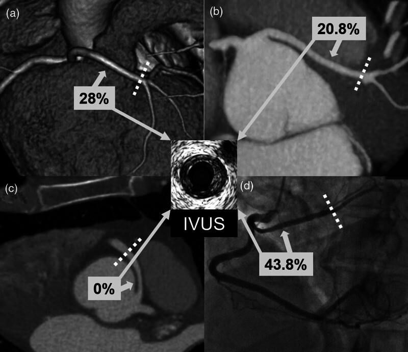
Fig. 3.
Illustration of CAD prevalence (%) in the proximal segment according to the type of AAOCA course. Panel A: CT image of a left main with a prepulmonic course; panel B: CT image of a left main with a subpulmonic course; panel C: CT image of a right coronary artery with an interarterial course; panel D: angiographic view of a circumflex artery with a retroaortic course. Dotted lines indicate the border between the proximal and distal segments. AAOCA, anomalous aortic origin of a coronary artery; CAD, coronary artery disease; CT, computed tomography; IVUS, intravascular ultrasound.

