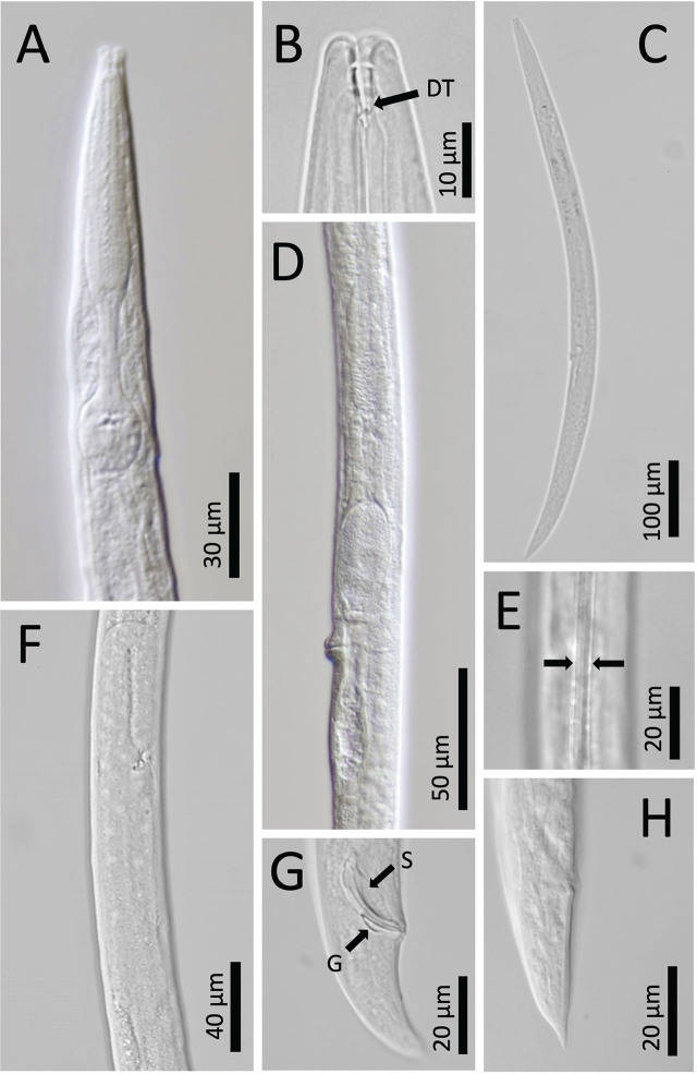
Figure 3:
Panagrolaimus namibiensis n. sp. light micrographs. A: Anterior body region and pharynx. B: Lip region, stoma, stegostomal dorsal tooth (DT). C: Full body, female. D: Part of reproductive tract, female. E: Lateral field. F: Part of testis, male. G: Posterior body end, spicule (S), gubernaculum (G), male. H: Posterior body end, rectum, female.

