Fig. 3.

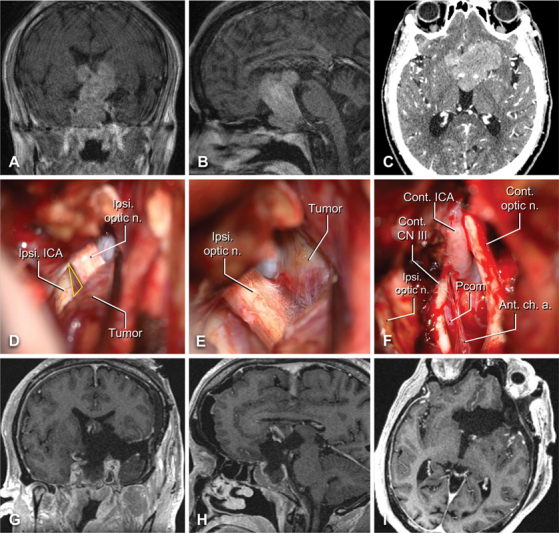
Illustrative case one – pituitary adenoma. Preoperative contrast-enhanced T1-weighted coronal ( A ) and sagittal ( B ) MRI slices are shown alongside axial CT angiogram ( C ). Intraoperative images demonstrating the optico-carotid triangle ( D ), yellow triangle), the ipsilateral optic nerve with tumor attached ( E ), and the contralateral anatomical structures after tumor removal ( F ) . ( G–I ) Comparable postoperative contrast-enhanced T1-weighted MRI demonstrating resection of the lesion with minor residual in the sphenoid sinus and right cavernous sinus. Abbreviations: AChA, anterior choroidal artery; CN, cranial nerve; cont., contralateral; ICA, internal carotid artery; ipsi, ipsilateral; optic n., optic nerve; PCoA, posterior communicating artery.
