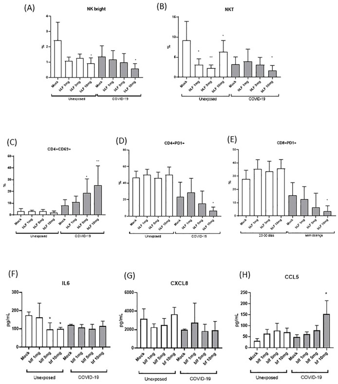
Figure 3.
In vitro effects of bLf on subpopulations of NK and T cells from unexposed and COVID-19 groups. (A) Percentage of NKbright (CD3−CD56highCD16−), (B) NKT(CD3+CD56+), (C) CD4+ activated T cells (CD69+), (D) exhausted CD4+ T cells (PD-1+), and (E) exhausted CD8+ T (PD-1+) cells upon stimulation with different bLf concentrations (1, 5, and 10 mg/mL), as well as unstimulated (mock). Levels of (F) IL-6, (G) CXCL8 (IL-8), and (H) CCL5 (RANTES) detected in supernatant obtained from cell stimulation conditions. *p < 0.05 and **p < 0.01.

