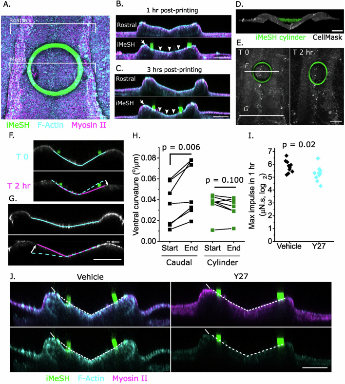
Extended Data Fig. 10. iMeSH cylinders do not disrupt tissue morphology and allow to quantify actomyosin-dependent pro-closure impulse.
a. Dorsoventral confocal image showing F-actin and myosin-II wholemount staining of a chick embryo with an iMeSH cylinder between the neural folds. Note this also illustrates the robust attachment of iMeSH to tissues, withstanding washes in solution and repositioning for imaging. Brackets through the iMeSH and 100 μm rostral to it indicate the positions of optical crosssections in B-C. b, c. Optical cross-sections through actomyosin-stained embryos fixed 1 hour or 3 hours following iMeSH printing. Arrowheads indicate persistent apical F-actin enrichment, arrows indicate normal morphology of the neural fold tips lateral to the iMeSH. In A-C: images are representative of 3 independent embryos. Scale bars = 100 μm. d. 3D reconstruction showing an iMeSH cylinder suspended between the RNP neural folds. Scale bar = 100 μm. e. Projections showing cylinder deformation over two hours. F and G indicate the positions of the corresponding panels. Scale bar = 100 μm. f, g. Optically-resliced projections showing the curvature of the ventral neuroepithelium (annotated) below the neural fold tips. Dashed lines indicate the T0 apical contour, and the white arrows indicate the change in bending. Scale bar = 50 μm. h. Quantification of the ventral neuroepithelial curvature at the start of imaging and 1.5-2 hours later in seven independent embryos caudal to, or at the level of, the cylinder. P values by twotailed T-test paired by embryo. i. Impulse was calculated as the maximum force applied to the i3D cylinder within 1 hour. Points represent independent embryos. Log2-normalised values of vehicle embryos are compared against Y27-treated embryos by t two-tailed test, n = 10 per group. j. Optical cross-sections through the neuroepithelium of wholemount stained vehicle and ROCKinhibited embryos with iMeSH cylinders showing diminished F-actin in the latter. Scale bar = 50 μm. Images are representative of 3 independent embryos per treatment group.

