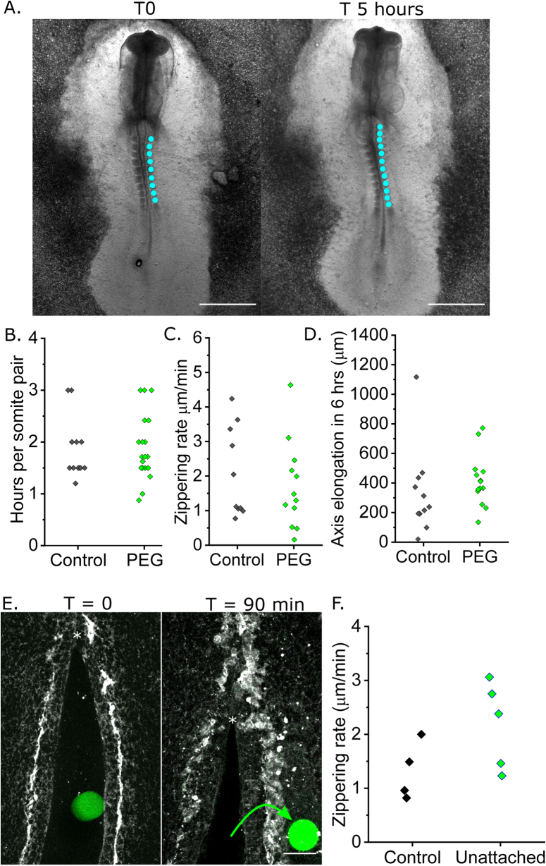
Extended Data Fig. 1. i3D polymer and printing do not diminish embryo development.
a. Brightfield images of a chick embryo in EC culture at T0 and T 5 hours. Cyan dots indicate somites. Scale = 500 μm. b-d. Quantification of parameters to compare growth between control embryos in EC culture and those treated with 30% HCC PEG liquid polymer. Points represent independent embryos. B. Rate of somite gain, n = 13 (control) and n = 21 (PEG). C. Caudal zippering point progression relative to a somite landmark, n = 10 (control) and n = 12 (PEG). D. Embryo axis elongation, n = 12 (control) and n = 15 (PEG). e. Sequential confocal images of a chick embryo posterior neuropore soon after i3D printing a pillar between its neural folds but not attached to its tissues, and 90 minutes later when theNunattached cylinder had been extruded from the neuropore (green arrow). * indicates thezippering point, scale bar = 50 μm. f. Quantification of rate of zippering point progression in the posterior neuropores of control embryos live-imaged without i3D printing (N = 4) and in embryos with unattached pillars printed inside their neuropore lumen (N = 5). Points represent individual embryos.

