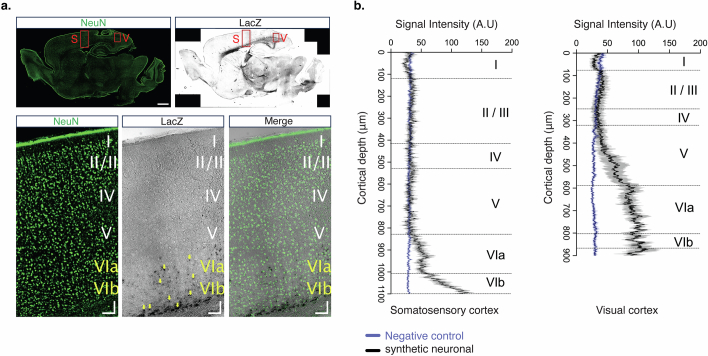
Extended Data Fig. 10. Immunohistochemistry of N1 CRE activity in the mouse cortex.
(a) Representative fluorescence and brightfield images showing expression patterns of neuronal marker, NeuN (top left) and LacZ (top right) across the whole brain. Boxed regions represent the somatosensory cortex (S) and visual cortex (V), digitally zoomed in bottom image; scale bars: 1 mm (top images) and 100 µm (bottom images). Yellow arrows indicate LacZ expression in layer 6. (b) Fluorescence intensity profile plots from quantification of LacZ signal intensity across layers in the somatosensory cortex and visual cortex for non-transgenic control (blue) and N1 CRE transgenic mouse (black).

