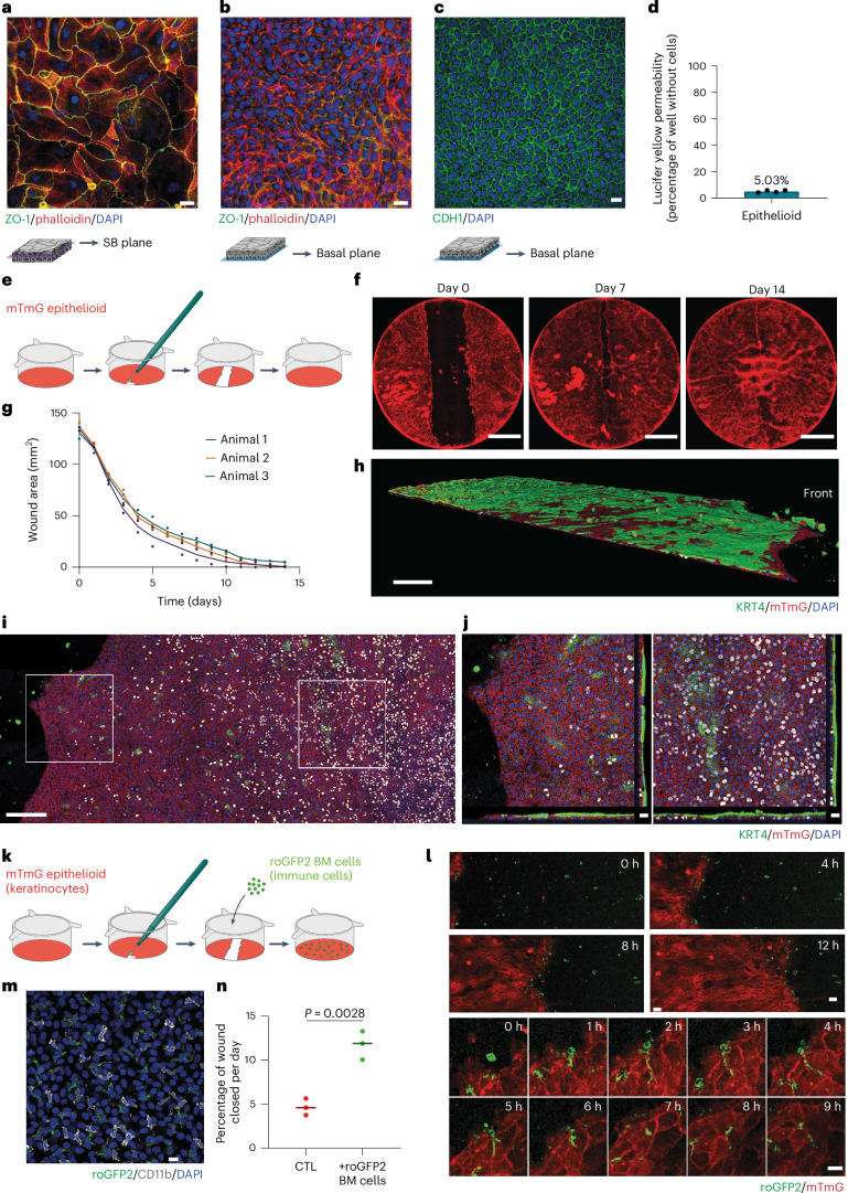
Fig. 4. Epithelioids have barrier function and repair capacity.
a,b, Esophageal epithelioids grown in mFAD immunostained with TJP1 (ZO-1) antibody (tight junctions, green), phalloidin (actin, red) and DAPI (nuclei, blue). Suprabasal (a) and basal layer (b) planes selected from the same culture area. Scale bar, 20 µm. Images are representative of three biological replicates. c, mFAD-grown esophageal epithelioids immunostained for CDH1 (adherens junctions, green) and DAPI (nuclei, blue); the basal layer plane is shown. Scale bar, 20 µm. d, Lucifer yellow permeability assay. Lucifer yellow is added for 30 min to the upper culture compartment of esophageal epithelioids and its transference to the lower compartment is quantified and compared with inserts without cells (100% permeability). n = 8 inserts from 4 mice. Each dot represents the average permeability of the inserts from each mouse. e–g, Esophageal epithelioids established from Rosa26mTmG mice and incubated in mFAD were wounded using a microscalpel (e). Daily images were taken in an Incucyte system (f) and the wound area was quantified (g). Each dot corresponds to a different culture, their color indicates the mouse of origin. Lines connect means of cultures from the same mouse. n = 6 inserts from 3 mice. Scale bar, 5 mm. h–j, Immunostaining of a Rosa26mTmG insert during the wound healing process using KRT4 (green), membrane Tomato (red) and DAPI (blue). h, Rendered confocal z-stack of a portion of the wound healing culture. 3D scale bar, 200 µm. i, Left: basal layer plane of a z-stack with white squares selecting a front area and a rear area of the wound. Orthogonal sections of the front (i, middle) and rear (j, right) areas selected from the left-hand panel. k–n, Rosa26mTmG esophageal epithelioids cultured in mFAD were wounded as in e, with the addition of bone marrow cells extracted from Rosa26mito-roGFP2-Orp1 mice (green) to the upper compartment right after wounding. k, Protocol scheme. l, Confocal live imaging images showing cell front and immune cells during wound healing (upper) and magnification of the cell front to follow immune cell internalization in the membrane Tomato membrane GFP (mTmG) epithelial cell layer (lower). Scale bar, 20 µm. m, Immunostaining of CD11b (gray) in an esophageal epithelioid co-cultured with bone marrow derived Rosa26mito-roGFP2-Orp1 cells (green), DAPI (blue). Scale bar, 14 µm. n, Quantification of the proportion of wound closed per day. Unpaired two-tailed Student’s t-test. Lines represent mean values. n = 3 biological replicates for each condition. BM, bone marrow; CTL, control; roGFP2, reduction-oxidation sensitive green fluorescent protein 2; SB, supra basal.

