Fig. 1.
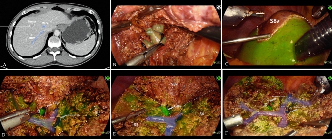
The novel ICG and HSA complex combined with “ negative-staining” fluorescence-guided laparoscopic anatomical sub-segmentectomy. A Preoperative enhanced computed tomography revealed that the tumor was situated in the ventral segment of segment 8 (S8v). B The ventral branch of segment VIII (G8v) was identified and occluded. C After successful negative staining, the fluorescent demarcation line between S8v and the parenchyma intended for preservation was highly distinct and conspicuous. D-E After the complete separation of S8v from segments 5 and 4A, the root of G8v was visibly exposed in the surgical field. F Only two ligation clips, which were used to ligate G8v and the vein draining S8v were visible in the surgical area. Additionally, the vein draining segment 4A, the middle hepatic vein, and the intersegmental vein were all clearly exposed in this cross-section

