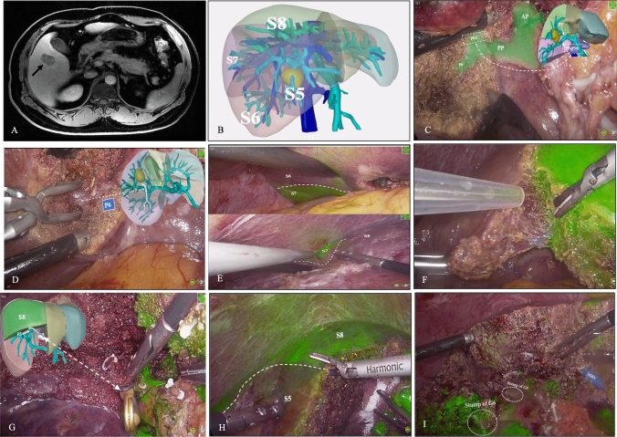
Fig. 3.
A A 44-year-old man with hepatitis B virus-related cirrhosis presented with a hypervascular nodule at the junction of hepatic segments 5 and 6 (black arrow) on magnetic resonance imaging which was highly suspicious for hepatocellular carcinoma. B A three-dimensional reconstruction showed the tumor was closely attached to branches G5 and G6 and was located between hepatic veins V5 and V6. C The pedicle of segment 6 (P6) and the dorsal pedicle of segment 5 (PPa) were exposed. D The PPa was transected and the anterior pedicle and P6 were isolated. E–F ICG-HSA was injected intravenously using an infusion pump for the first negative-staining procedure. Clear fluorescent demarcation was observed. G) The pedicle supplying dorsal segment 5 was dissected and occluded using a bulldog clamp. After that, IGC-HSA was injected a second time. H Fluorescent demarcation between segments 8 and 5 was observed. I An en-bloc specimen containing segments 5 and 6 was successfully resected. The middle hepatic vein and stump of G6/G5 were exposed on the resection surface

