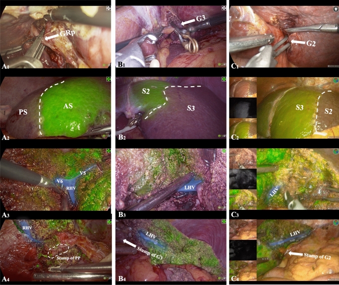
Fig. 4.
A Laparoscopic right posterior segmentectomy: the extrahepatic Glissonean pedicle of the right posterior section (GRp) was clamped using a bulldog clamp (A1). After intravenous injection of ICG-HSA, clear fluorescent demarcation was observed between the posterior section (PS) and anterior section (AS) (A2). The landmark hepatic veins (V5, V6, and the right hepatic vein [RHV[) between the PS and AS were precisely located on the demarcation line (A3). After completion of the surgery, the entire course of the stump of the Posterior Pedicel (PP) and the RHV were visible (A4). B & C Laparoscopic hepatectomy of segments 2/3: the extrahepatic Glissonean pedicle of segments 2/3 (G3/G2) was clamped using a bulldog clamp (B1/C1). After intravenous injection of ICG-HSA, clear fluorescent demarcation was observed between S2 and S3 (B2/C2). The left hepatic vein (LHV) between S2 and S3 was precisely located on the demarcation line (B3/C3). After completion of the surgery, the entire course of the stump of G3/G2 and the LHV were visible (B4/C4)

