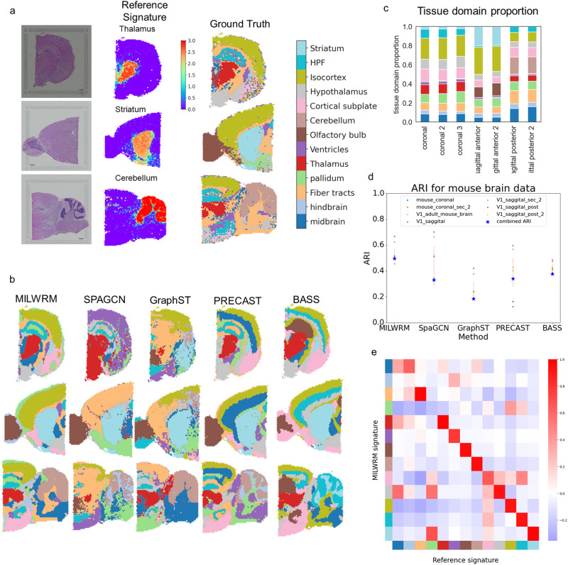
Fig. 4. MILWRM detects consensus tissue domains in ST data from different mouse brain cross-sections.
a Reference signature (middle—thalamus, striatum, and cerebellum top to bottom, respectively) based ground truth annotation (right) in mouse brain ST data (scale bar = 500 µm). b MILWRM, SpaGCN, GraphST, PRECAST, and BASS detected tissue domains (k = 13) overlaid on three mouse brain ST samples. c Proportion of tissue domains in slides each bar corresponding to the legend on the left. d Scatter plot for ARI and consensus ARI. e Correlation matrix for overall correlation between MILWRM and reference scores for each TD and anatomical region across all spots.

