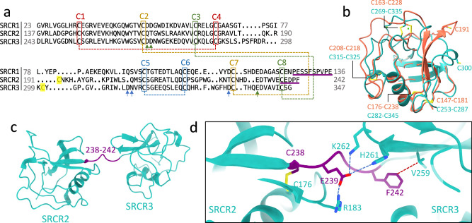
Fig. 2. SRCR domains of AIM.
a Structure-based sequence alignment of the three SRCR domains of AIM. SRCR2 and SRCR3 are cryo-EM structures and SRCR1 is the AF2 predicted model. The four conserved disulfide bonds are highlighted with dash lines with different colours. The unpaired cysteine in SRCR2 and SRCR3 are in yellow. The three green arrows indicate the residues of calcium binding site 1 in SRCR3 which are also conserved in SRCR1. The three blue arrows point at the calcium binding site 2 residues in SRCR3. The sequences of linkers between SRCR1 and SRCR2 and between SRCR2 and SRCR3 are underlined in purple. b Superposition of SRCR2 (red) and SRCR3 (cyan) cryo-EM models showing the four conserved disulfide bonds and the two unpaired cysteine residues (C191 and C300). c AIM model from the complex with IgM showing linker (highlighted in purple) between SRCR2 and SRCR3 domains. d Zoomed-in view of the linker-domain interactions with potential salt bridges as blue dashed lines and van der Waals contact as red dashed line.

