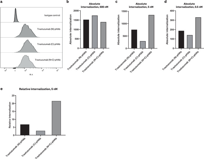
Fig. 4. Internalization of pair-FORCE-generated HER2 antibodies in different formats on HER2-expressing SK-BR-3 cells.
Internalization of Trastuzumab-derived HER2 binders in the N, C, and N + C format by HER2-expressing SK-BR-3 cells was assessed by flow cytometry using HER2 antibodies conjugated with the pH-sensitive dye pHAb. The conjugated HER2 antibodies were generated with pair-FORCE technology. The fluorescence of the pHAb dye is poor at neutral pH but increases substantially upon trafficking into the acidic environment of the endo-/lysosomal pathway13. For all panels, n = 1 independent replicates were performed. a Fluorescence histogram from flow cytometry experiments of SK-BR-3 cells treated with pHAb-conjugated HER2 binders at a saturating concentration of 500 nM. pHAb fluorescence was measured in the PE (phycoerythrin) channel. b The same experiment as (a), but absolute internalization is depicted as a bar graph. Absolute internalization is defined as the geometric mean of pHAb fluorescence in flow cytometry experiments. c Absolute internalization of pHAb-conjugated HER2 antibodies as in (b), but at a concentration of 5 nM. d Absolute internalization of pHAb-conjugated HER2 antibodies as in (b, c), but at a concentration of 0.5 nM. e Relative internalization of Trastuzumab-derived HER2 antibodies at a concentration of 5 nM. Relative internalization is calculated as the ratio of absolute internalization to absolute binding, which is derived from flow cytometry binding experiments with AF488-labeled HER2 antibodies generated by pair-FORCE (see Methods section for details). The absolute binding is defined as the geometric mean of AF488 fluorescence of SK-BR-3 cells treated with 200 nM of AF488-conjugated HER2 antibodies. N, C, and N + C refer to the binder formats in Fig. 1b. Source data for Fig. 4b–e are provided as a Source Data file.

