
Fig. 1.
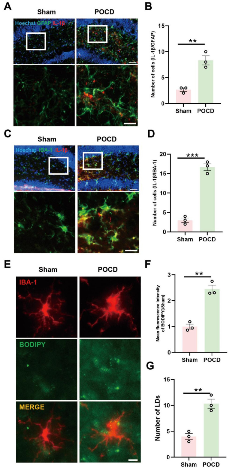
Microglia-mediated neuroinflammation and LDs accumulation in the hippocampus of mice with postoperative cognitive impairment. (A) Co-labeling of astrocytic glial fibrillary acidic protein (GFAP) and inflammatory cytokine IL-1β in the hippocampal region of mice (scale bar = 40–20 μm); quantification of co-labeled cell count in (B) (n = 3). (C Co-labeling of microglia marker IBA-1 and inflammatory cytokine IL-1β in the hippocampal region of mice (scale bar = 40–20 μm); quantification of co-labeled cell count in (D) (n = 3). (E Immunofluorescence staining of hippocampal microglia cells, with BODIPY probe marking LDs in the hippocampal area (scale bar = 10 μm). Fluorescence intensity of BODIPY in (F) and quantification of labeled LDs in (G) (n = 3). *P < 0.05, **P < 0.01, ***P < 0.001 compared with the corresponding group, as determined by t test.