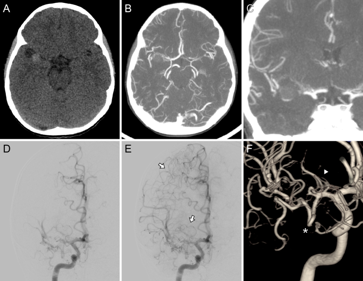
FIG. 1.
Preoperative imaging. Axial NCCT scan (A) showed a slightly hyperdense ovoid lesion in the right sylvian cistern. Axial (B) and coronal (C) CTA demonstrated an MCA occlusion at the level of the distal end of the M1 segment, although its distal branches were present. Digital subtraction angiography after right ICA injection, posteroanterior view, early (D) and late (E) arterial time, and three-dimensional (3D) rendering (F). This angiographic study revealed the lesion’s specific location and a lumen tapering before it, suggestive of an occlusive dissecting aneurysm. Distal flow reconstitution was noted through leptomeningeal collaterals originating from the ACA and from an anastomotic network involving deep perforators (white arrows, E). Additionally, the lenticulostriate arteries (white arrowhead, F) and an early temporal branch (white asterisk) originated proximal to the lesion.

