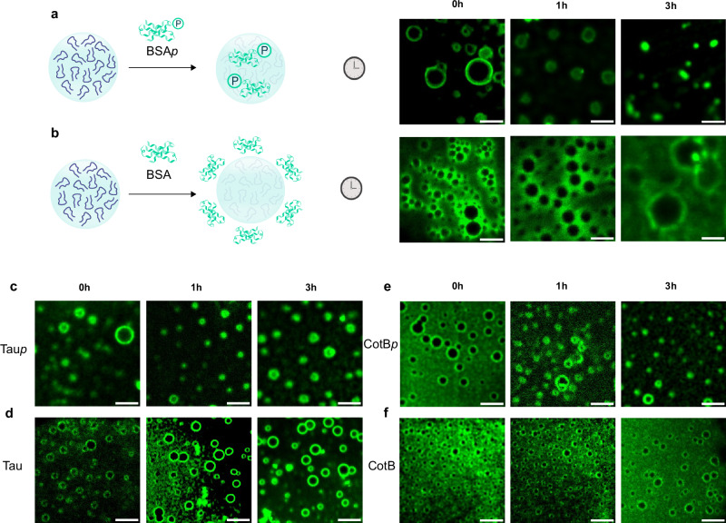
Fig. 4. Affinity-mediated molecular uptake within P7 coacervates.
Time-lapse microscopy captures of the uptake of phosphorylated assemblies inside P7 coacervates. FITC-labeled BSAp and BSA, Alexa-labeled Taup and Tau, and Alexa-labeled CotBp and CotB were incubated with P7 coacervates for 3 h. Representative fluorescence confocal images highlight the molecular uptake within P7 coacervates (a) for FITC-labeled BSAp (c) Alexa-labeled Taup (e) Alexa-labeled CotBp and the exclusion of (b) BSA, (d) Tau, and (f) CotB at the boundaries of P7 coacervates. Scale bar set to 10μm. Full field of view in Supplementary Figs. 8–12.

