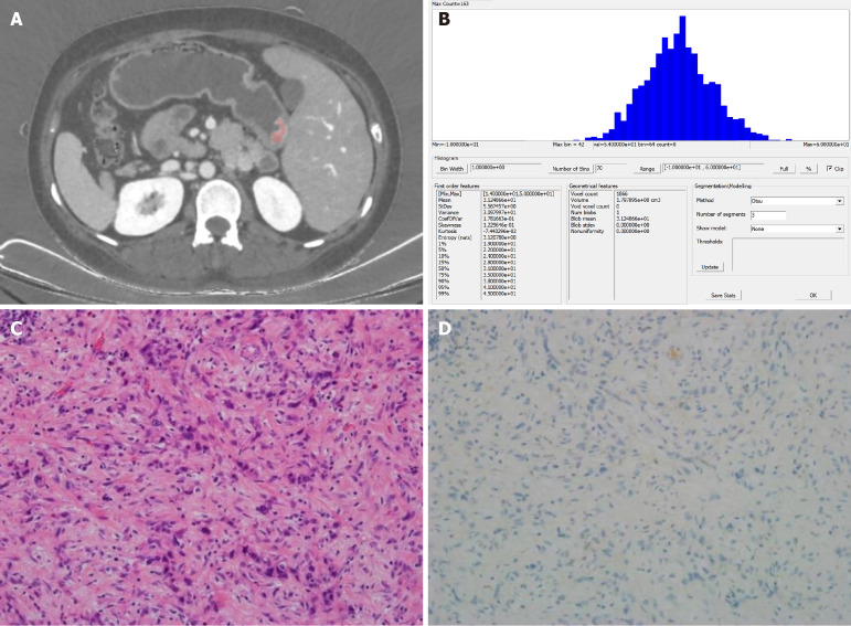
Figure 3.
Images from a 43-year-old woman with poorly differentiated gastric adenocarcinoma. A: Portal venous-phase spectral computed tomography image showing clear (pink) enhancement of the lesion, located at the gastric antrum; B: Histogram of parameter distributions for the whole tumor (minimum = 14.000, maximum = 50.000, mean = 31.249, standard deviation = 5.567, skewness = 0.123, kurtosis = 0.074, 1st-99th percentiles = 19.000, 22.000, 24.000, 28.000, 31.000, 35.000, 38.000, 41.000, and 45.000, respectively); C and D: Microscopic pathological (HE staining, 200 ×) and immunohistochemical images, respectively, showing a poorly differentiated adenocarcinoma with a Lauren classification of diffuse type, vascular and neural invasion, and negative HER2 staining.

