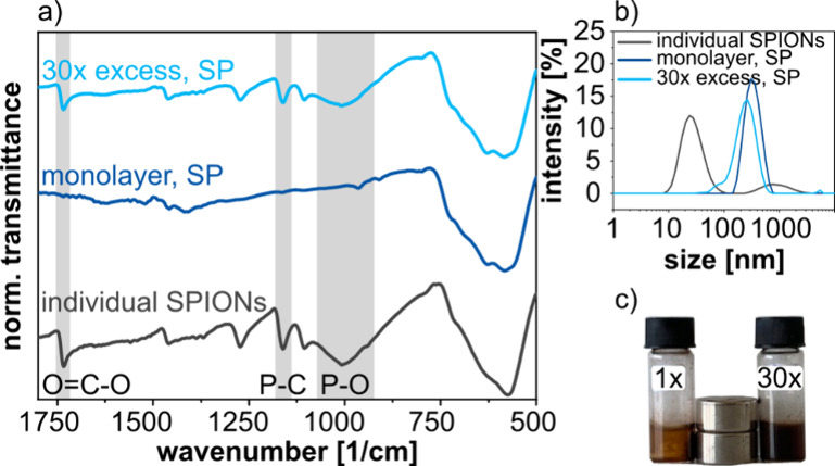
Figure 3.

(a) FTIR spectra of individual SPIONs (edge length ∼ 14 nm) after surface modification with BiB-UDPA ligands (black curve), SPs based on 12 nm cubic SPIONs treated with an amount of BiB-UDPA ligands corresponding to roughly a monolayer coverage of the SPs (dark blue line), and of SPs treated with a 30-fold higher concentration of BiB-UDPA (light blue line). (b) Corresponding DLS size distributions of individual SPIONs and SPs after the reaction with BiB-UDPA corresponding to either a monolayer or a 30-fold higher concentration. (c) Photography of the SP dispersions with 1× and 30× concentration of BiB-UDPA under the influence of two NdFeB disk magnets (N42).54 The concentration of SPs in both samples was identical (10 mg/mL).
