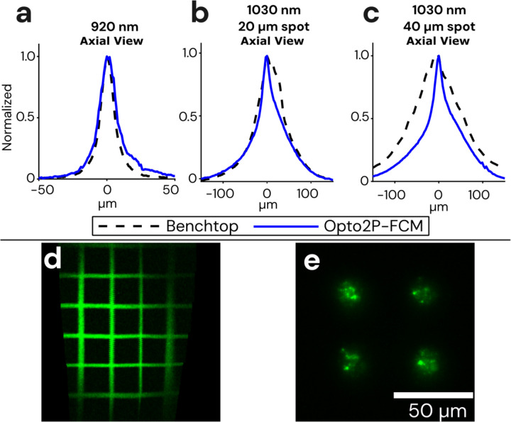
Figure 2: Optical resolution and field of view measurements of the Opto2P-FCM.
(a) Axial resolution of the Opto2P-FCM was characterized by measuring the 2P fluorescence signal of a uniform flat fluorescent target scanned through the focus of the 920 nm laser (blue solid line, 14 μm FWHM) compared with a benchtop two-photon microscope with a 10x/0.4 NA objective (dashed line, 11 μm FWHM). Axial resolution of the 1030 nm patterned photostimulation was measured for a (b) 20 μm diameter circle using the Opto2P-FCM (blue solid line, 50 μm FWHM) compared to the benchtop microscope (dashed line, 54 μm FWHM) and for a (c) 40 μm diameter circle using the Opto2P-FCM (blue solid line, 54 μm FWHM) compared to the benchtop microscope (dashed line, 124 μm FWHM). (d) Opto2P-FCM image of a 50 μm grid slide showing a field of view of ~200 μm × 300 μm. (e) Image of photostimulation pattern (4 circles with 20 μm diameter) at the focus of the Opto2P-FCM recorded by 2P fluorescence from a thin rhodamine sample.

