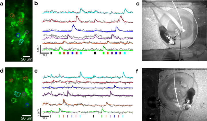
Figure 3: Demonstration of cell-specific 2P patterned photostimulation and simultaneous 2P imaging in a freely moving mouse.
The stimulation sequence consists of all ROIs together followed by stimulation of each ROI individually. Top panels were taken using a 0.5 m length CFB in visual cortex, with 5 ms/frame of stimulation for 2 seconds, bottom panels were taken using a 1 m length CFB in somatosensory cortex, with 10 ms/frame of stimulation for 700 ms. (a) and (d) show the fields and targeted/detected ROIs, with solid line circular ROIs indicating the targeted stimulation regions and dashed line ROIs indicating those detected by CaImAn. (b) and (e) show ΔF/F traces of jGCaMP7s activity during the recording timelapse color matched to ROIs in (a) and (d). Solid lines represent ΔF/F denoised traces from CaImAn and overlaid dashed lines are raw ΔF/F traces of the stimulation ROI. Color matched bars at the bottom represent stimulation times and durations, with black representing the stimulation of all ROIs. (c) and (f) show a still frame of the mouse during the recording. Behavioral video is shown in Video S1.

