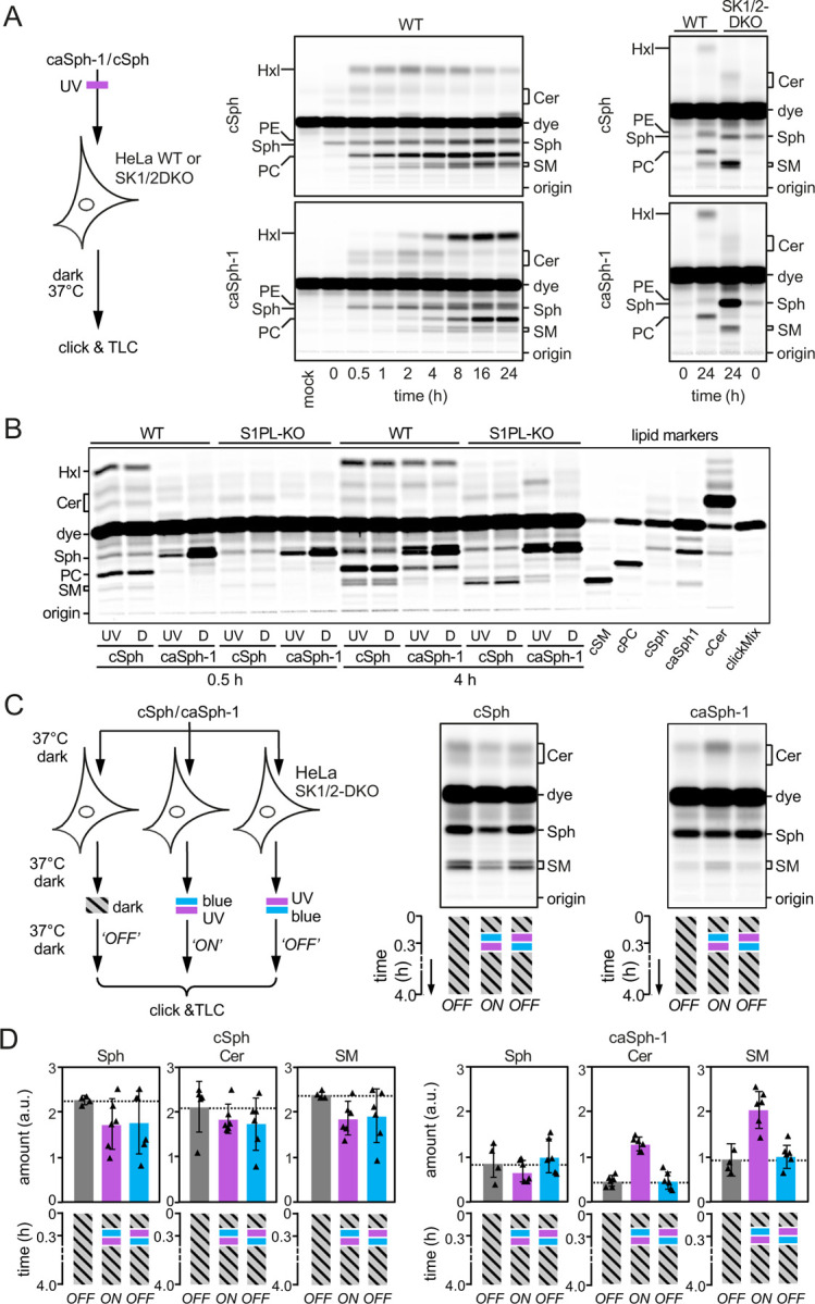
Figure 3. caSph-1 enables optical control of sphingolipid biosynthesis in living cells.

(A) HeLa wild-type (WT) or SK1/2DKO cells were cultured in the presence of UV-irradiated cSph or caSph-1 for the indicated period of time. Metabolic conversion of cSph and caSph-1 was monitored by TLC analysis of total lipid extracts click-reacted with Alexa-647. (B) HeLa wild-type (WT) or S1PL-KO cells were cultured in the presence of UV-irradiated (UV) or dark-adapted (D) cSph or caSph-1 for 0.5h or 4h. Metabolic conversion of cSph and caSph-1 was monitored as in (A). (C) HeLa SK1/2DKO cells were cultured in the presence of dark-adapted cSph or caSph-1 for 30 min, irradiated with blue- followed by UV-light or vice-versa, and then incubated for up to 4h in the dark. Cells kept in the dark throughout the incubation period served as control. Metabolic conversion of cSph and caSph-1 was monitored as in (A). (D) Quantification of clickable sphingosine (Sph), ceramides (Cer) and sphingomyelin (SM) levels in cells treated as in (C). Data shown are mean values ± s.d. from six biological replicates (n = 6).
