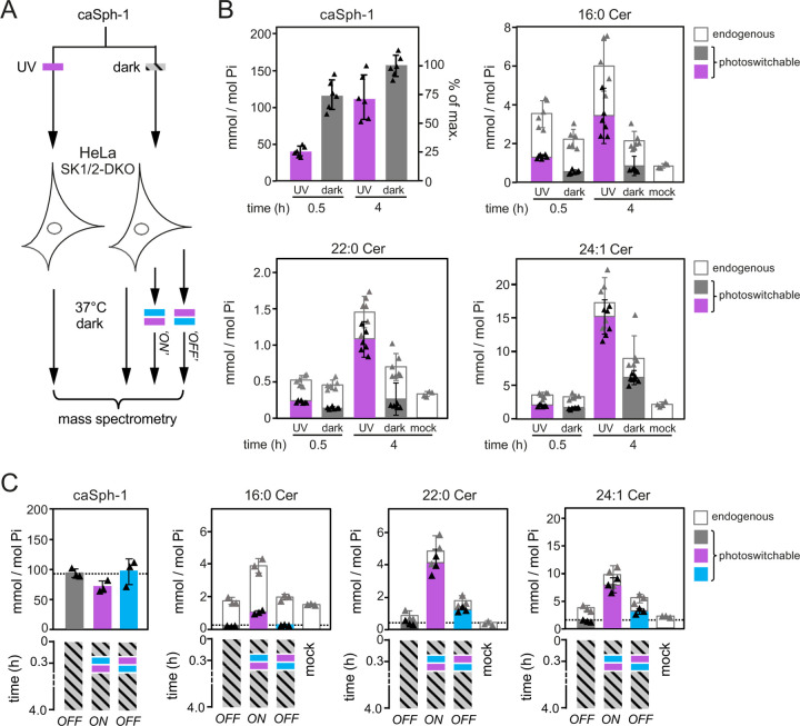
Figure 4. Optical manipulation of cellular ceramide pools.
(A) Schematic outline of lipid mass spectrometry on HeLa SK1/2DKO cells incubated with dark-adapted or UV-irradiated caSph-1 and subjected to distinct light-regimes. (B) HeLa SK1/2DKO cells were fed dark-adapted or UV-irradiated caSph-1, incubated in the dark for 0.5 or 4 h and then subjected to targeted quantitative lipid mass spectrometry to determine cellular levels of caSph-1, photoswitchable and endogenous Cer species. Values are plotted as mole fraction of total phospholipid (left-hand vertical axis) and % of total input (for caSph-1, right-hand axis). Data shown are mean values ± s.d. from six biological replicates (n = 6). (C) HeLa SK1/2DKO cells were incubated with dark-adapted caSph-1 for 30 min, washed, irratiated with blue- followed by UV-light or vice versa and then incubated for up to 4h in the dark. Cells kept in the dark throughout the incubation period served as control. Cellular levels of caSph-1, photoswitchable and endogenous Cer species were quantified as in (B). Data shown are mean values ± s.d. from six biological replicates (n = 6).

