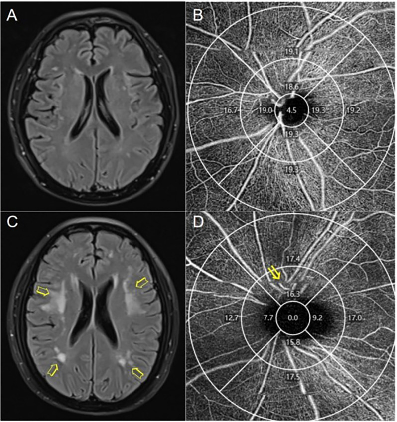
Fig 1. Representative images of OCT-A and brain MRI from study participants.

(A) Mild white matter lesions (Fazekas scale score = 1, Scheltens scale score = 7). (B) Peripapillary vessel density (VD) of a patient with mild white matter lesions. (C) Severe white matter lesions (Fazekas scale score = 6, Scheltens scale score = 25). (D) Patient with severe WMH (arrows) showing decreased peripapillary vascular density in the superior quadrant of the inner ring (arrows).
