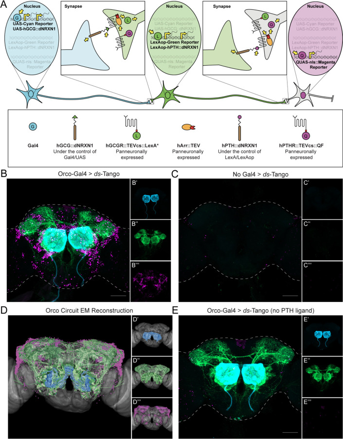
Figure 2. Design and implementation of ds-Tango in the olfactory system.
(A) Schematic and components of ds-Tango. In flies carrying a Gal4 driver, the presynaptic reporter (cyan) and the GCG ligand (hGCG::dNRXN1) are expressed in the starter neurons. The GCG ligand localizes to the presynaptic sites of the starter neurons and activates the GCGR Tango fusion (hGCGR::TEVcs::LexA*) across the synapse on the monosynaptic partners. Upon activation of the GCGR, the hArr::TEV fusion protein is recruited to it, TEV cleaves its recognition site (TEVcs) releasing LexA*. LexA* then translocates to the nucleus and initiates the expression of the PTH ligand (hPTH::dNRXN1) and of the monosynaptic reporter (green). The PTH ligand localizes to the presynaptic sites of the monosynaptic partners and activates the PTHR Tango fusion (hPTHR::TEVcs::QF) across the synapse on the disynaptic connections. Upon activation of the PTHR, the hArr::TEV fusion protein is recruited to it, TEV cleaves its recognition site (TEVcs), releasing QF. QF then translocates to the nucleus and initiates the expression of the nuclear disynaptic reporter (magenta). The various steps in the process are indicated by yellow arrows.
(B) Driving ds-Tango in the peripheral olfactory system using Orco-Gal4 labels OSNs (cyan, shown in B’), their monosynaptic partners LNs and OPNs (green, shown in B’’), and the nuclei of their disynaptic connections (magenta, shown in B’’’).
(C) A brain of a control fly bearing the ds-Tango components, but no Gal4 driver exhibits no background neurons in the presynaptic channel (cyan, shown in C’), virtually no background neurons in the monosynaptic channel (green, shown in C’’), and the nuclei of a few background neurons in the disynaptic channel (magenta, shown in C’’’).
(D) EM reconstruction of the Orco circuit projected on a template brain (gray) reveals OSNs (cyan, shown in D’), LNs and OPNs (green, shown in D’’), and the nuclei of third-order olfactory neurons (magenta, shown in D’’’).
(E) A brain of a control fly bearing Orco-Gal4 and a version of ds-Tango lacking the PTH ligand exhibits labeling in OSNs (cyan, shown in E’), LNs and OPNs (green, shown in E’’), but no staining of disynaptic partners except for the nuclei of a few background neurons (magenta, shown in E’’’).
Maximum intensity Z-stack projection of whole-mount brains are shown in B, D, and E. Dashed lines in B, D, and E depict the approximate outline of the fly brains. Scale bars = 50 μm.

