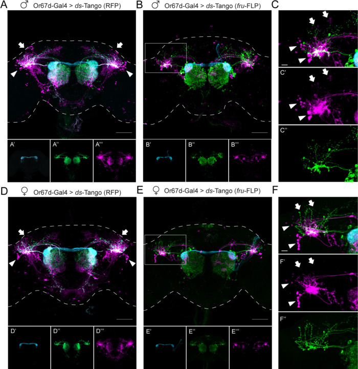
Figure 4. ds-Tango reveals sexual dimorphism in the lateral horn in the Or67d circuit.
(A) Driving ds-Tango with Or67d-Gal4 in males labels Or67d-OSNs (cyan, shown in A’), their monosynaptic partner LNs and OPNs (green, shown in A’’), and their disynaptic connections (magenta, shown in A’’’). Arrows indicate the dorsal cluster neurons; arrowheads indicate the lateral cluster neurons.
(B) Driving ds-Tango with Or67d-Gal4 and genetically restricting disynaptic reporter expression to fru-FLP+ neurons in males labels Or67d-OSNs (cyan, shown in B’), their monosynaptic partner LNs and OPNs (green, shown in B’’), and their fru-FLP+ disynaptic connections (magenta, shown in B’’’).
(C) A higher magnification image of the gray inset in (B) highlighting the left LH reveals PNs targeting the ventral region of the LH (green, shown in C’’), overlapping with the neurites of fru-FLP+ disynaptic connections (magenta, shown in C’). Note the presence of lateral cluster neurons (arrowheads) and the dorsal cluster neurons (arrows) in the male brain.
(D) Driving ds-Tango with Or67d-Gal4 in females labels Or67d-OSNs (cyan, shown in D’), their monosynaptic partner LNs and OPNs (green, shown in D’’), and their disynaptic connections (magenta, shown in D’’’). Arrowheads indicate the lateral cluster neurons; arrows indicate the location of dorsal cluster neurons.
Note the presence of lateral cluster neurons in both the male (arrowheads in A) and female (arrowheads in D) brains and the prominent dorsal cluster neurons in the male (arrows in A) brain that are less prominent or absent in the female (arrows in D) brain.
(E) Driving ds-Tango with Or67d-Gal4 and genetically restricting disynaptic reporter expression to fru-FLP+ neurons in females labels Or67d-OSNs (cyan, shown in E’), their monosynaptic partners LNs and OPNs (green, shown in E’’), and their fru-FLP+ disynaptic connections (magenta, shown in E’’’).
(F) A higher magnification image of the gray inset in (E) showing the left LH reveals PNs targeting the ventral region of the LH (green, shown in F’’), overlapping with the neurites of fru-FLP+ disynaptic connections (magenta, shown in F’). Note the presence of lateral cluster neurons (arrowheads) and the absence of dorsal cluster neurons (arrows) in the female brain.
Maximum intensity Z-stack projection of whole-mount brains are shown in A, B, D, and E. Dashed lines in A, B, D, and E depict the approximate outline of the fly brains. Scale bars, 50 μm.

