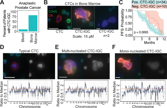
Figure 1.
Large tumor cells are found in BM of late-stage prostate cancer patients. (A) Enumeration of patients with matched blood and bone marrow samples with at least 1 CTC-IGC present in liquid biopsy. (B) Representative images of CTC and CTC-IGC found in BM aspirate. Scale bars set to 15 μM. (C) PFS from patients with or without CTC-IGC found in BM samples. (D) Representative image of typical CTC found in BM with merged and DAPI channels (top) and its genomic copy number profile (bottom). (E) Representative image of CTC-IGC found in bone marrow with merged and DAPI images (top) and its genomic copy number profile (bottom). (F) Representative image of mono-nucleated CTC-IGC found in bone marrow with merged and DAPI images (top) and its genomic copy number profile (bottom).

