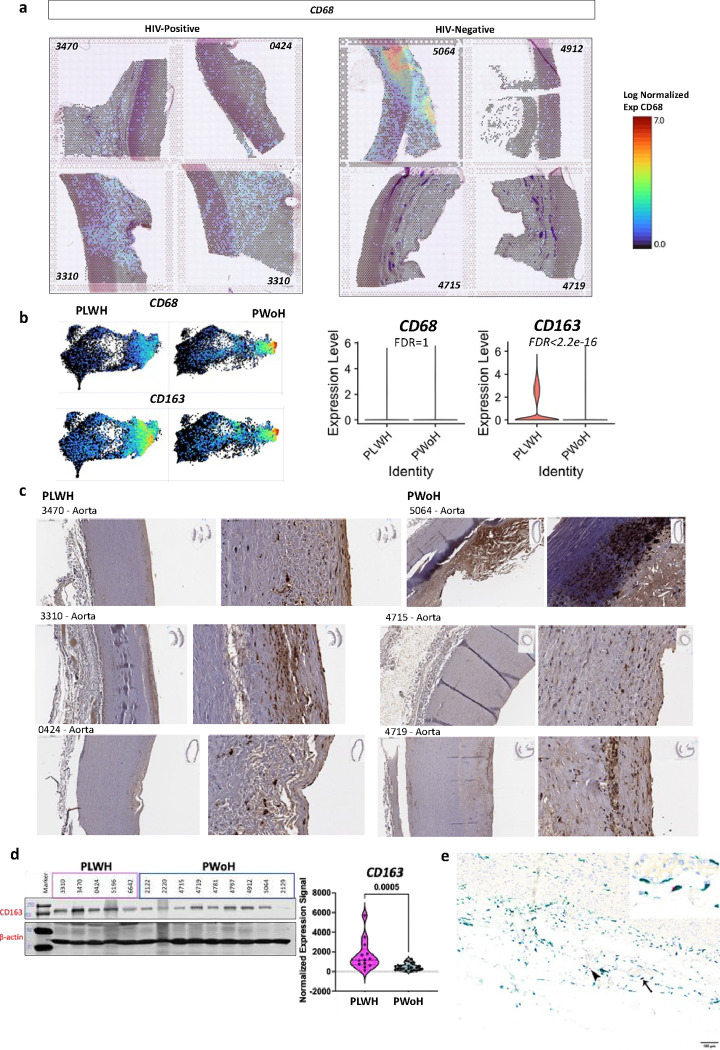
Figure 3. CD163 is higher in the aorta from PLWH compared to PWoH and colocalizes with HIV RNA.
a, Thoracic aorta obtained from deceased donors were stained with H&E stains and processed for Visium spatial gene expression analysis. The log normalized expression of CD68 is shown with a spatial resolution. b, The combined HIV-positive and HIV-negative aorta are projected to a UMAP representing each spot that captures data (55μm per spot). Both CD68 and CD163 expression as shown on the UMAPs. Violin plots show the log-normalized expression of CD68 and CD163 in all eight samples by HIV status. c, IHC, and d, western blot showing CD163 protein expression. Violin plots show CD163 expression normalized to the housekeeping proteins β-actin from 3 separate runs. e, Dual detection of CD163 protein expression by immunohistochemistry (green) and HIV RNA hybridization (red) within the aortic adventitia. HIV signals were detected adjacent to (arrowhead) or within CD163-labeled macrophages (thin arrow, inset).

