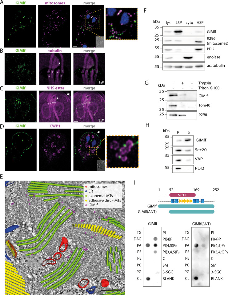
Fig 2. GiMlf is localized in the vicinity of mitosomes, nucleation zones of cytoskeletal components, and other membrane-bound compartments.
(A) Localization of endogenously BAP-tagged GiMlf in Giardia using confocal microscopy. The cells were stained with an anti-BAP antibody (green), an anti-GL50803_9296 antibody (mitosomal marker; magenta), nuclei stained with DAPI (blue). DIC image of the corresponding cell is shown in the corner of the merged image, scale bar: 2 μm. (B,C) Localization of GiMlf in trophozoites using expansion microscopy (ExM), 3.7 expansion factor. Scale bars: 4 μm. (B) The cells were stained with an anti-GiMlf antibody (green) and an anti-acetylated tubulin antibody (magenta) or (C) anti-BAP antibody (green) and NHS ester dye that labels primary amines of proteins. The basal bodies are marked by asterisks, the dense band is indicated by an arrowhead and the disc margin is marked by an arrow. (D) Localization of GiMlf in encysting cells (48 h post induction) using confocal microscopy. The cells were stained with anti-CWP1 antibody (green) and anti-BAP antibody (magenta), nuclei stained with DAPI (blue). The disc margin is marked by an arrow. The DIC image of the corresponding cell is shown in the corner of the merged image, scale bar: 2 μm. (E) Electron tomography of the central region between two Giardia nuclei depicting immunogold-labeled endogenously V5-tagged GiMlf and reconstructed subcellular structures. The image shows the presence of GiMlf (magenta dots) at the base of the basal bodies (green), adhesive disc microtubules (yellow) and at the mitosomes (red), scale bar: 250 nm. (F) Western blot of Giardia cellular fractions labeled with anti-GiMlf antibody and compartment-specific antibodies. Lysate (lys), low-speed pellet (LSP; containing cytoskeleton and nuclei), cytoplasm (cyto) and high-speed pellet (HSP; containing membrane-bound organelles), 9296—mitosomal marker protein (GL50803_9296) of unknown function, PDI2 –ER marker–protein disulfide isomerase 2, enolase–cytosolic marker, ac. tubulin–acetylated tubulin. (G) Trypsin treatment of the combined LSP and HSP fractions in the presence or absence of 1% Triton X-100. Tom40 and GL50803_9296 were used as markers for protease-accessible and membrane-protected proteins, respectively. (H) The combined fractions of LSP and HSP were treated by sodium carbonate to release peripherally associated proteins from cellular membranes. S–fraction of proteins released into the supernatant, P–fraction of proteins pelleted together with membranes. Sec20, VAP, and PDI2 were used as markers for integral membrane proteins. (I) The membrane lipid strip assay demonstrates the affinity of GiMlf for signaling components within the membrane. Recombinant GiMlf was detected using an anti-GiMlf antibody. TG–triglyceride, DAG–diacylglycerol, PA–phosphatidic acid, PS–phosphatidylserine, PE–phosphatidylethanolamine, PC–phosphatidylcholine, PG–phosphatidylglycerol, CL–cardiolipin, PI–phosphatidylinositol, C–cholesterol, SM–sphingomyelin, 3-SGC– 3-sulfogalactosylceramide, PI(4)P–phosphatidylinositol (4)-phosphate, PI(4,5)P2 –phosphatidylinositol (4,5)-bisphosphate, PI(3,4,5)P3 –phosphatidylinositol (3,4,5)-trisphosphate.

