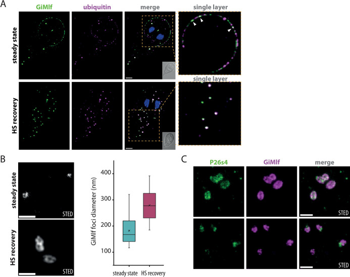
Fig 5. GiMlf-P26S4 foci are ubiquitin-rich bodies that enlarge after heat shock.
(A) Localization of GiMlf and ubiquitin in G. intestinalis using confocal microscopy in steady state and after 4 hours of recovery after heat shock. The cells were stained with anti-BAP (green) and anti-ubiquitin (magenta) antibodies. The enlarged images show a single layer of the image stack. The arrows point to locations on the disc margin, where ubiquitin and GiMlf colocalize. Nucleic DNA was stained with DAPI (blue). DIC image of the corresponding cell is shown in the corner of the merged image. Scale bars: 2 μm. (B) Comparison of GiMlf foci before and after heat shock and 4 hours of recovery using STED microscopy. The cells were stained with anti-BAP antibody. Scale bars: 0.5 μm. Box plots compare the foci diameters measured at its widest point in each state (n = 30; significant difference determined with the two-tailed t-test with equal variance, P-value = 1.72xE-9). (C) Localization of GiMlf and V5-tagged P26s4 after heat shock and 4 hours of recovery using STED microscopy. The cells were stained with anti-BAP (green) and anti-V5 (magenta) antibodies. Scale bars: 0.5 μm.

