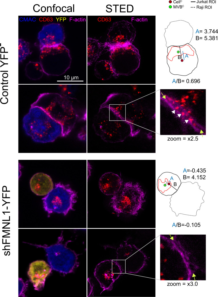
Figure 10. STED image of CD63+ nanovesicles at the synaptic cleft.
C3 control clone cells untrasfected or expressing shFMNL1-HA-YFP were challenged with CMAC-labeled SEE-pulsed Raji cells (blue) for 1 hr, fixed, stained with phalloidin AF647 (magenta) and anti-CD63 (red) and imaged simultaneously by confocal and STED microscopy. Two representative control YFP- and FMNL1-interfering (shFMNL1-HA-YFP) Jurkat cells forming immune synapse (IS) with Raji cells are shown. Confocal (left) and STED (right) optical sections are shown, and enlarged (2.5× and 3× zoom) views of the IS areas are shown in the right side. The yellow arrows on the enlarged IS images label the edges of the synaptic cleft, which is the narrow, lane-shaped space between the two cells enclosed by the two F-actin-rich (magenta) plasma membrane leaflets. CD63+ multivesicular bodies (MVB) from the Jurkat cells are located nearby to the IS and some CD63+ nanovesicles (white arrows) are located at the synaptic cleft in the control YFP- example. The diagrams used to calculate the MVB polarization index (PI) data in both cell groups are represented in the right side of the images. The zoom of the indicated regions of interest (ROIs) (white square) is included below the diagrams. The percentage of synaptic conjugates on which was evident the presence of CD63+ nanovesicles at the synaptic cleft was 17% for control YFP-, and 0% for FMNL1-interfering (shFMNL1-HA-YFP), respectively, with at least 35 synapses analyzed per condition. Images are representative of the data from several independent experiments (n=3) with similar results.

