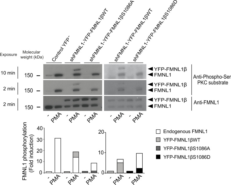
Figure 3. S1086 in FMNL1β is phosphorylated upon protein kinase C (PKC) activation.
C3 control clone was unstransfected (Control YFP-) or transfected with either FMNL1-interfering expressing interference-resistant YFP-FMNL1βWT (shFMNL1-HA-YFP-FMNL1βWT), YFP-FMNL1βS1086A, or YFP-FMNL1βS1086D constructs. Subsequently, cells were stimulated or not (-) with PKCδ activator phorbol myristate acetate (PMA) for 30 min. The different cell groups were lysed and immunoprecipitated with anti-FMNL1. These immunoprecipitates (IPs) were analyzed by western blot (WB), first with anti-Phospho-Ser PKC substrate antibody (two different expositions) and then reprobed with anti-FMNL1 to normalize Phospho-Ser PKC substrate signal for FMNL1 protein levels. The lower graph represents the normalized fold induction of phosphorylation of the different FMNL1 variants. Results are representative of data from several independent experiments (n=3) with similar results.

