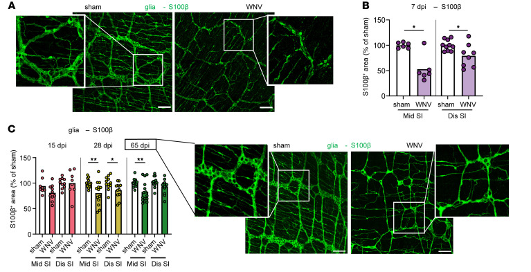
Figure 2. WNV infection affects enteric glial networks.
(A–C) The muscularis externa was isolated from middle and distal regions of the small intestine of sham- or WNV-infected C57BL/6J mice at (A and B) 7 dpi or (C) 15, 28, and 65 dpi and stained for glia (S100β). The fraction of area that stained positive for S100β was determined, and the values were normalized to sham-infected mice. Representative images show S100β staining in the middle region of the small intestine in sham- and WNV-infected mice at (A) 7 dpi or (C) 65 dpi. (A and C) Scale bars: 100 μm. Original magnification, ×2.5 (enlarged insets). Data were pooled from (A and B) 2 experiments (n = 5–10) and (C) (left to right) 2, 3, and 4 experiments (n = 6–10, 12–13, and 13–16). Column heights indicate the mean values. *P < 0.05 and **P < 0.01, by 2-tailed Mann-Whitney U test.

