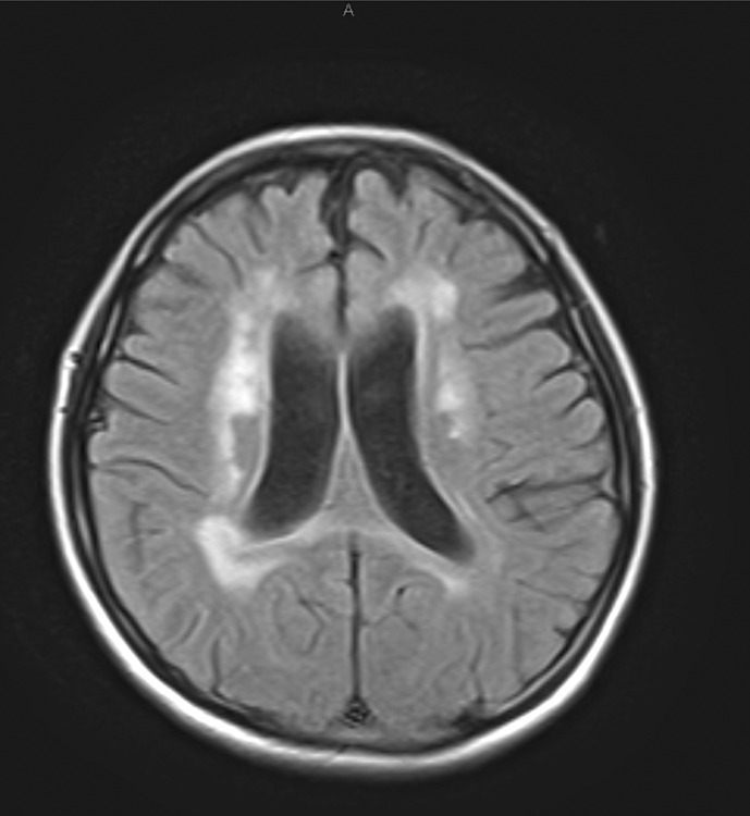
Fig. 1.
Cerebral magnetic resonance imaging (FLAIR) performed at the patientʼs first visit revealing extensive, patchy, and confluent hyperintensities in the periventricular and deep white matter. Compared to age, the enlargement of the body from the anterior horn of the ventricle on both sides and front-cortical atrophy was remarkable.

