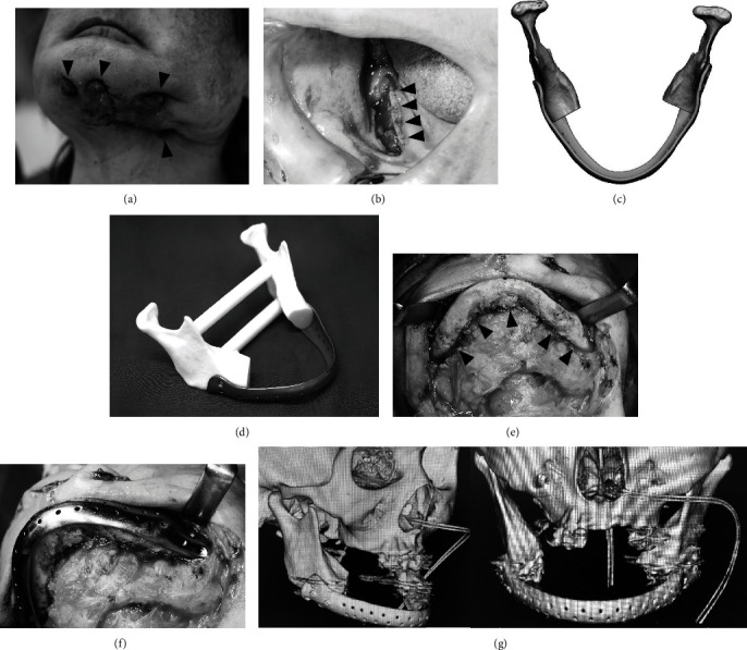
Figure 2.

Case 2. (a) Preoperative extraoral photograph. Four large fistulas are observed extending from the chin to the left mandible (arrowhead). (b) Preoperative intraoral photograph. Mandibular bone exposure is observed in the right lower gingiva (arrowhead). (c) Three-dimensional (3D) design of the reconstruction plate. (d) 3D model and reconstruction plate. (e) Intraoperative photograph. The mandible bone is clearly indicated (arrowhead). (f) Confirming the plate on the mandible bone. (g) Postoperative computed tomography. The adaptation between the plate and bone was acceptable.
