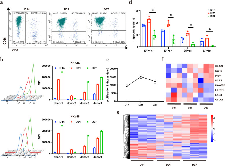
Fig. 1.
Peripheral blood derived natural killer cells (NK) were gradually activated by a feeder-free system in vitro. a The percentage of NK cells (CD3−CD56+) cultured in vitro at day 14, 21 and 27 was detected by using Flow cytometry. b Mean fluorescence intensity (MFI) of surface receptors NKp44 and NKp46 at different time points during NK cell expansion in vitro. c Growth curve of NK cells. d Comparison of killing activity of NK cells against HCT116 cells with different ratio of effector to target at different expansion time. e Heat map of differentially expressed genes. f Differentially expressed activating and inhibitory receptors of NK cells. One-way ANOVA was used for comparisons between multiple groups. All results are presented as the mean ± SD (n = 3)

