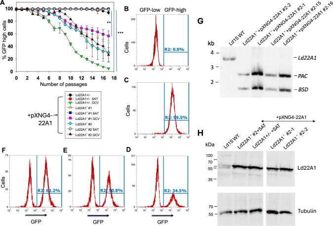
Fig. 4. CYP5122A1 is indispensable during the promastigote stage.
A Promastigotes were continuously cultivated in the presence or absence of ganciclovir (GCV) or nourseothricin (SAT) and passed every three days. Percentages of GFP-high cells were determined for every passage. Symbols and error bars represent the means and standard deviations from three independent repeats. Two-tailed ANOVA without adjustment (**p < 0.01, ***p < 0.001). Source data are provided as a Source Data file. After 14 passages, WT (B) and Ld22A1−+pXNG4-22A1 #2 parasites grown in the presence of SAT (C) or GCV (D) were analyzed by flow cytometry to determine the percentages of GFP-high cells (indicated by R2 in the histograms). Two clones (E: #2-1; F: #2-2) were isolated from the GFP-low population in D by FACS followed by serial dilution and amplification in the presence of GCV and analyzed for GFP expression by flow cytometry. G To verify the presence of episomal CYP5122A1 allele, single clones isolated from the GFP-low (#2-1 and #2-2) and GFP-high (#2-15 and #2-16) populations of Ld22A1−+pXNG4-22A1 #2 parasites (D) were expanded in the presence of GCV and examined by Southern blot using a flanking region probe upstream of the CYP5122A1 ORF probe. Bands corresponding to endogenous CYP5122A1, episomal CYP5122A1, and antibiotic resistance markers PAC/BSD were indicated. H To examine the CYP5122A1 protein levels, whole cell lysates from log phase promastigotes before (Ld22A1− + pXNG4-22A1 #2 and Ld22A1 + /- +pXNG4-22A1) and after (#2-1 and #2-2) sorting were analyzed by Western blot using anti-LdCYP5122A1 (top) or anti-α-tubulin antibodies.

