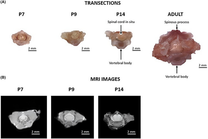
FIGURE 1.

(A) Cross sections of perfused surgically removed thoracic vertebra 10 for postnatal day P7, P9, and P14, and 9‐week (adult) Sprague–Dawley (SD) rats and (B) cross sections of ex vivo spinal columns with the spinal cord in situ imaged using MRI (magnetic resonance imaging) for postnatal days P7, P9, and P14 SD rats.
