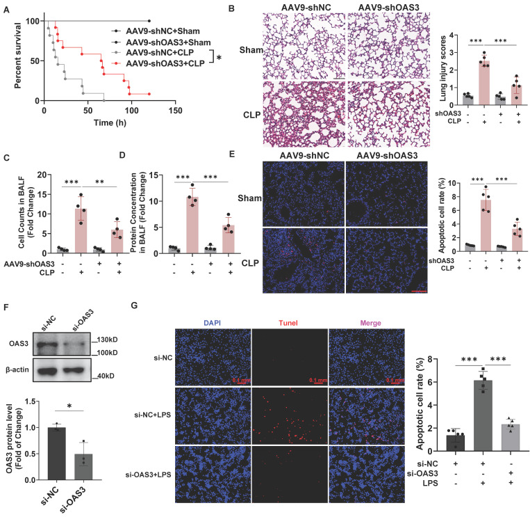
Figure 3.
OAS3 accounts for sepsis-induced acute lung injury and lung epithelial cell apoptosis. (A) Kaplan-Meier survival analysis of Sham and CLP mice intratracheally injected with AAV9-shNC (negative control) or AAV9-shOAS3 (n=11). (B) Representative H&E staining and histological scores of lung sections in Sham and CLP mice intratracheally injected with NC-shRNA or OAS3-shRNA AAV9 (n=5, scale bar: 100 μm). (C) The cell number and (D) protein concentration in bronchoalveolar lavage fluid (BALF) of Sham and CLP mice intratracheally injected with NC-shRNA or OAS3-shRNA AAV9 (n=4). (E) TUNEL-positive cells (stained with Cy3, red) and nuclei (stained with DAPI, blue) in lung tissue of Sham and CLP mice intratracheally injected with NC-shRNA or OAS3-shRNA AAV9 (n=5, scale bar: 100 μm). (F) OAS3 protein level in MLE12 cells transfected with specific siRNA targeting OAS3 (n=3). (G) TUNEL-positive cells in MLE12 cells transfected with OAS3 siRNA followed by LPS treatment were observed and analyzed using a fluorescence microscope (Zeiss) (n=5, scale bar: 100 μm). Data are shown as Mean ± SD. *P<0.05, **P<0.01, ***P<0.001.

