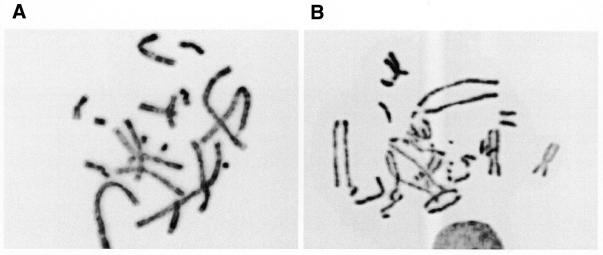
Figure 6.
Impaired sister chromatid cohesion. Shown are representative pictures of (A) metaphase spread from a V79B cell with normal cohesion and (B) metaphase spread from a CL-V4B cell with impaired sister chromatid cohesion (see Table 4). Both metaphase spreads were generated after treatment with colcemid.

