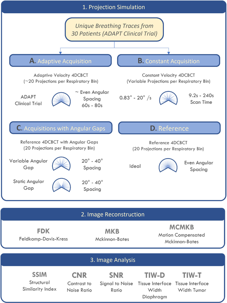
FIGURE 1.
Summary of the study workflow. Patient and simulated respiratory data were separated into 10 respiratory phases and a XCAT volume was generated for each phase. Image reconstruction was performed using FDK, MKB, and MCMKB algorithms and images were analyzed using SSIM, CNR, SNR, TIW-D, and TIW-T.

