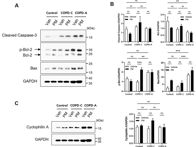
Fig. 5.
Cell damage mechanisms. (A) Western blot analysis was performed to detect expression of apoptosis related proteins in lung tissues from each group with GAPDH as a loading control, (B) Results of densitometry analysis. Protein levels of cleaved caspase-3, Bcl-2, phosphorylated Bcl-2, and Bax were quantified and normalized to GAPDH band intensity, (C) Necrotic protein cyclophilin A expression was assessed and quantified. Bcl-2, B-cell lymphoma protein 2; Bax, Bcl-2 associated X; GAPDH, glyceraldehyde-3-phosphate dehydrogenase; PM, particulate matter. **: p < 0.01 and ***: p < 0.001, ns: non-significant

