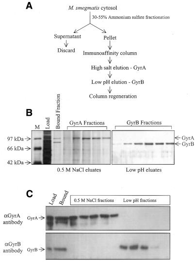
Figure 1.
Immunoaffinity purification of M.smegmatis DNA gyrase. (A) Schematic representation of the purification protocol detailed in Materials and Methods. (B) SDS–PAGE and silver staining of the affinity-purified fractions. M represents size markers with the indicated molecular masses. (C) Western blot analysis of eluted fractions probed with GyrA- and GyrB-specific antibodies. Load and bound represent the protein loaded on and bound to the column, respectively.

