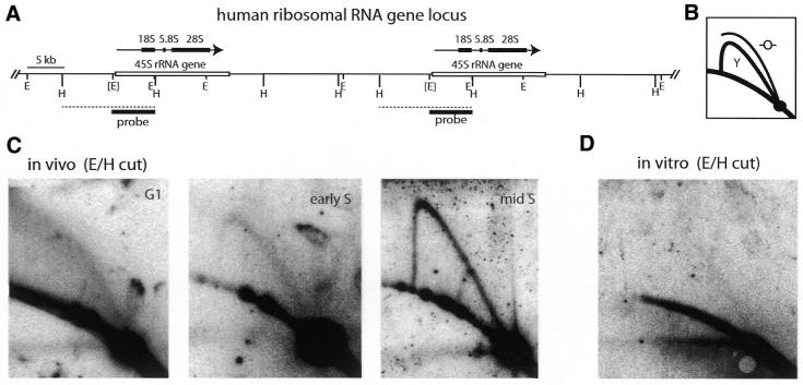
Figure 4.
Lack of initiation of DNA replication at the human rDNA locus in early S phase in vivo and in vitro. (A) Map of the human rDNA locus (cf. 9,14). Two of the repeated rDNA transcription units and the intergenic spacers are shown. The region coding for the 45S rRNA precursor is indicated by a white box, the 45S rRNA transcript as a black arrow with the 18S, 5.8S and 28S rRNA coding sequences highlighted as a series of black boxes. Restriction sites for EcoRI (E) and HindIII (H) are shown. The EcoRI site 5′ to the transcription start site is polymorphic and is indicated by brackets. The position of the hybridisation probe (EcoRI fragment B; 9) is indicated by a black box and the position of the analysed polymorphic E/H DNA fragment is indicated by a dashed line. (B) Principle of neutral/neutral 2D gel electrophoresis mapping of replication intermediates (46). DNA restriction fragments containing replication intermediates are first run from top to bottom and in the second dimension from left to right. Linear fragments migrate along the lower arc and unit length fragments accumulate at a discrete spot on this arc. Restriction fragments containing a single replication fork migrate on arc Y and fragments containg a centrally located defined origin of bidirectional replication migrate on arc O. (C) Neutral/neutral 2D gel analysis of rDNA replication in vivo. Total genomic DNA was purified from cells in G1 phase (arrested by mimosine), early S phase (released from mimosine for 3 h) and mid S phase (released from a double thymidine block for 3 h), enriched for replicative intermediates, cut with EcoRI and HindIII and subjected to 2D gel electrophoresis. Replicative intermediates were visualised by hybridisation to radioactively labelled EcoRI fragment B and autoradiography. (D) 2D gel analysis of rDNA replication in vitro. G1 phase nuclei from mimosine-arrested cells were incubated in cytosolic extract from proliferating cells for 3 h. Replicative intermediates were isolated and visualised as detailed for (C).

