. 2024 Oct 24;12(10):e009416. doi: 10.1136/jitc-2024-009416

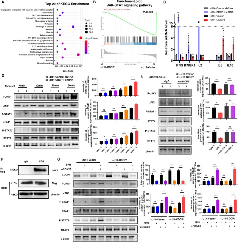
Figure 5. CDCP1 inhibits the JAK-STATs signaling of T cells. (A) Kyoto Encyclopedia of Genes and Genomes (KEGG) analysis of RNA-seq data of active T cells co-cultured with of CDCP1 overexpressing U14 cell vs control U14 cells for 24 hours. (B) GSEA analysis of the differentially expressed genes (from RNA-seq datasets) involved in the suppression of JAK-STAT signaling pathway in T cells co-cultured with CDCP1 overexpressing U14 cells compared with T cells co-cultured with control U14 cells. p<0.001. (C) qPCR validation of the expression of genes downstream JAK-STAT signaling pathway in active T cells co-cultured with different CDCP1 expression level U14 cells. (D) The activation of the JAK-STAT pathway was assessed in the lysates of T cells co-cultured with control and CDCP1 knockdown U14 cells, following stimulation with anti-CD3 and anti-CD28 antibodies for 10–30 min. (E) The activation of the JAK-STAT pathway was assessed in the lysates of T cells co-cultured with control and CDCP1-overexpressing U14 cells, with or without anti-CD6 blockade. (F) Immunoprecipitation assay identified the interaction between CD6 and JAK1 in T cells. Murine T cell were transfected with CD6-3xFlag plasmid. Cell lysates were immunoprecipitated with Flag antibody and analyzed by immunoblot with anti-JAK1 and anti-Flag. (G) CDCP1 inhibitor 8PN activates the JAK1-STAT1/3 signaling pathway in T cells. After pretreated with 8PN or DMSO for 24 hours, the activation of the JAK-STAT pathway was assessed in the lysates of T cells co-cultured with control and CDCP1-overexpressing U14 cells. Significance was determined using Student’s t-test. *p<0.05, **p<0.01, ***p<0.001, ****p<0.0001. ns, not significant.

Figure 5